Eyetrraction is a platform completely dedicated to the eyes and hence we certainly cannot exclude the understanding of the anatomy of the eye.
But before moving to the parts of the human eye and their functions, let us understand the placement of the eyes in our body.
On the front portion of the skull, below the forehead lies a formation of hollow amongst seven bones. This formation is known as Orbit. The eyeball is positioned in the orbit.
Orbit is a hard structure and hence question that rises is, “How does the eye get its movement in it?”
Well, nature has its way. There is a substantial amount of fat layered between the eye and the orbit to facilitate easy movement of the eye. However, the eye movement is supported by 6 muscles that are connected to the uppermost layer of the eye called the Sclera. We’ll learn more about this later on in this blog.
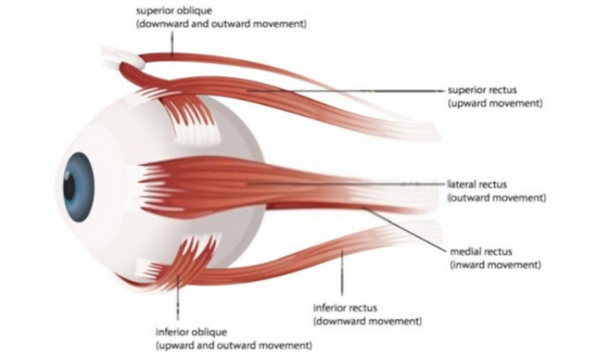
The six muscles also known as extraocular muscles are:
- Superior rectus
- Inferior rectus
- Medial rectus
- Lateral rectus
- Inferior oblique
- Superior oblique
At the back of the orbit remains a hole through optic nerves that build a connection between the eye and the brain.
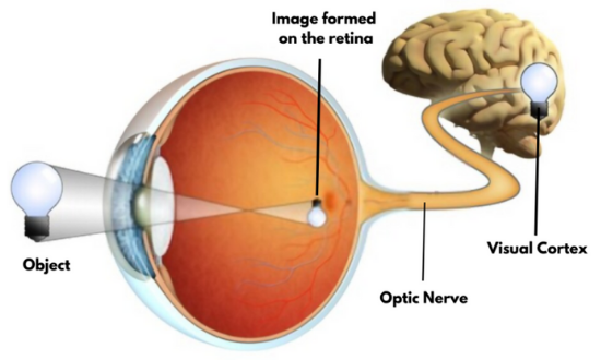
The connection between the eye and the brain is superfast. With each glance, our brain understands what it sees and takes impulsive decisions based on it. Sounds odd but it is true that it is actually the brain that sees. Eyes just act like the windows. To understand you need to learn the mechanism of the vision.
In short brief, light rays that travel into the eye form an inverted image on the retina which is then converted into electrophysiological impulses that are transmitted to the brain via Optic Nerves.
Two images are sent to the brain which is also known as Binocular Vision. This is because any image is viewed by each eye at two different angles. Hence the images are slightly different. The brain then merges these two images so as to understand the distance of the objects. This ability to sense distance is called Depth Perception.
The portion of the brain that receives the electrophysiological impulses, converts and interprets them is called the Visual Cortex.
Moving to the anatomy of the eye, let us start with the first most layer of the eye.
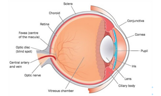
Cornea
The front-most part of the eye, the Cornea is a transparent covering of the eye having no color of its own. You can think of it as the glass layer of the watch that lets you see the interior parts such as the dial. Similar is the Cornea that acts as a cover for the internal parts such as the Iris and the lens.
The function of the Cornea is to focus the light rays the travel through its transparent layer. It is convex in nature and hence the light rays that passing through it converge to make their way into the eye through the pupillary gap.
The 12 mm in diameter, 0.5 mm in thickness and 8mm in curvature; Cornea is avascular by nature, meaning it has no blood supply. If the cornea had veins within it; the vision we would see would be like a grid view. The power of the cornea is +45 Dsph with a refractive index of 1.34.
The cornea is made of five layers; however, the surface is pores to get its supplement from the atmosphere, aqueous humor, and limbal blood.
Aqueous Humor
The clear liquid flowing between the Cornea and the Iris is the Aqueous Humor. The function of the ciliary body placed behind the Iris is to produce Aqueous humor. Around 0.5 ml of it is produced on daily basis.
This liquid is crucial to help maintain constant Intraocular pressure. In order to do so, it drains from the eye from tear ducts through the area called Drainage Angle. Along with that, it also supplies protein and nutrition to the cornea and the lens. Furthermore, it protects the eye from dirt.
Our eyes are designed by nature itself to protect them from external objects. Have you noticed? When a bug or small insect falls in your eye it dies immediately for two reasons: one is that you rub your eye vigorously & in defense, the bug releases some acids which are washed out by Watering Mechanism that pushes it into the corner from where it can be removed.
Even our eyelids facilitate this mechanism by working as wipers of the car that pushes all the dirt towards the lower lids from where it gets collected in the corner of the eye – canthus. This is called the Blinking Mechanism.
Having said that, to give readers an understanding of the complete water cycle of the eye, I have covered it in a different article altogether.
Pupil
It is nothing but a gap measuring about 3 to 7 mm in the Iris. The function of the Pupil is to control the amount of light coming into the eye.
Iris
Iris is often used in Biometrics since the color & design of Iris are unique for every human. No two people have the same design of the eye Iris.
The function of Iris is to control the size of the pupil. When it is dark your Iris contracts thus expanding the pupil for more light to enter the eye and when there is extra light it expands thus reducing the size of the pupil restricting excess light.
People can be seen with blue eyes, black eyes, brown, turquoise, and some other colors. It’s a beautiful creation of nature and a wonder in itself.
Wearing sunglasses excessively even in low light can reduce this natural functioning of the Iris. This needs to be taken care especially in the case of kids.
Crystalline lens
The lens is placed behind the Iris. It is absolutely transparent and also stretchable.
With that being true, it is also a fact that the lens is biconvex in nature. In other words, it is curved on both sides. Due to this, the power of the lens in an Emmetropic eye (normal) is +14.0 Dsph to +18.0 Dsph. It further results in the convergence of the light rays traversing through it to form a focal point on the retina. If there are any curvature differences or opacity in the lens, the patient finds difficulty in vision.
It is interesting how the lens functions. For viewing objects at a distance, it stretches and becomes thin at the center to facilitate more focusing of the picture on the retina. On the other hand, while viewing objects at a near distance, it contracts and thickens at the center for maximum focusing. When it comes thinner, the power of the lens reduces, and the focal length increases wherein when the lens thickens, its power increases and focal length reduces. This shrinking and the expanding mechanism is called Accommodation of the lens. It performs this mechanism with the help of Zonules. These are fibrous strands connecting the ciliary body and the lens. These are also known as suspensory ligaments that help in accommodation and also keep the lens in its place.
At the age of 38 – 40, this elasticity of the lens gradually reduces therefore, the near vision becomes weaker, the condition is known as Presbyopia Some people even experience this condition in their mid-thirties, known as Early Presbyopia which is mostly curable.
For near vision, plus power addition is required for its correction. In the early days before the invention of the progressive lens, people used bifocals, trifocals, Dbifocals for near vision correction. These were typical lenses divided into two parts with a visible line wherein the top part is for distance vision and the bottom part holds power for near vision. Trifocals also had the intermediate vision but the lines resulted in jump effects and later in their extinct use.
The progressive spectacle lens is largely adopted in modern times given its comfort. Multifocal lenses however are gradually gaining popularity.
The crystalline lens is made of protein and water. In most people around the age of 50, the lens becomes opaque, light cannot be transmitted and the vision becomes blurred or spotted. This condition is known as a cataract, treated by surgery.
In cataract surgery, the original opaque lens is removed and an IOL (Intraocular lens) is fitted instead. A small dissection is made at the limbus and the folded lens slides in which then opens up inside the eye and hooks to the ciliary body.
A major limitation of the IOL is that it’s not flexible hence accommodation is not achieved. Again correction is required for near vision.
By nature, the diameter of the lens is 9 – 10 mm, weight is 135mg, and the refractive index is 1.39.
Lens Capsule
The lens is wrapped in a transparent bag-like covering that holds the lens in its place. You can think of it as medical capsules holding medicinal powder within. During cataract surgery, the IOL is placed over the lens capsule to keep it from falling into the Vitreous Humor. We’ll be learning about this later in this article.
Sclera
The Sclera is the outermost layer of the eye. The previously mentioned 6 extraocular muscles are connected to the Sclera to facilitate easy movement of the eyeball. Furthermore, Sclera is a strong and opaque layer, what we see as the white part of the eye. Only 1/6th of the Sclera is visible from the front side of the eye.
The function of the Sclera is to protect the internal organs of the eye and helps in maintaining the basic shape of the eye. A thin layer of conjunctiva covers the sclera.
Choroid
It is the second layer after the sclera. This layer contains blood veins and its function is to give supplements to the eye and do the maintenance of the retina.
Retina
This is the third and most sensitive layer of the eye. Image focused through the lens is projected inverted on the retina. May this is what led to the invention of the camera!
The retina anatomy is interesting but a little complex. Let’s decode. The light rays focus on the centermost area of the retina. It is a structure designed for the most appropriate vision in any light.
As learned before, photoreceptors are present on the retina which converts the light rays into electrophysiological impulses. These are transmitted to the brain through optic nerves.
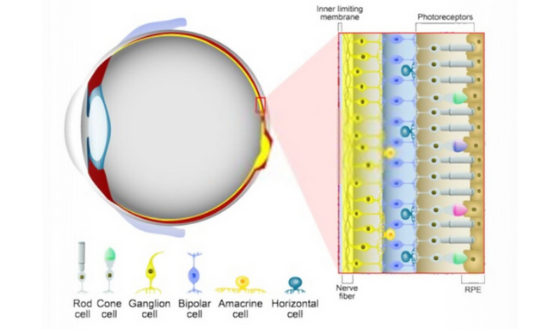
The photoreceptors are of two types: Rods are activated in the dark and only recognize black and white colors. Cones are activated in bright light & help in recognizing colors. The area where the cons are in maximum density is the macula.
The Macula is a structure for most sharp vision. But within macula lies fovea. The Fovea is the center of the macula with a high density of cones. Moving far from fovea i.e., towards the periphery of Macula, the density of cons reduces while rods increases.
It is been said that women’s eyes have more population of cones and hence can easily differentiate between two close colors.
Optic Nerves
As we know, the optic nerve is behind the eyeball. The function of the Optic Nerves is to transmit electrophysiological impulses to the brain. Each optic nerve from both the eye sends one image which is then merged by the brain in the visual cortex as explained before in the diagram. The good health of optic nerves is a crux matter.
“Glaucoma is a disease that is often associated with the elevated ocular pressure, in which damage to the optic nerve can lead to loss of vision and even blindness eventually.”
Optic Disc
All the layers of the retina end at a 1.5 mm pink-colored disc. There are no rods or cones present here. In other words, there is no vision in this area. The optic nerve starts here. This is also popularly known as the Blindspot.
Vitreous Humor
It is a translucent jelly-like blackish substance between the lens and the retina, forming the posterior portion of the eye. 80% of the eyeball is Vitreous Humor. It maintains the positioning of the parts of the eye, supports the retina, and helps in maintaining the Intraocular pressure.
“Due to the deposit of protein and blood cells for a longer period of time, clumps are formed in the vitreous humor. They cast shadows on the retina when they come in the path of light rays in the eye. These are known as Floaters.”
Lacrimal gland
This gland sits inside the orbit under the outside edge of the eyebrow. It produces water/ tears that form the tear film. The film itself is made of three layers – The mucous layer, the watery layer, and the oil layer. In some cases when the gland is weak, the patient suffers from dry eyes. Excess water gets drained from the tear duct present on the corner of the lower eyelid.
Meibomian gland
These are present under the lower eyelid responsible for producing oil for the tear film.
Sure, the anatomy of the eye is quite overwhelming but having known the complexity of the structure and the parts of the eye, taking good care of the eye cannot be overlooked.